What is the spinal cord?
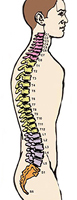
Between the body and the brain the spinal cord is the most significant structure. The spinal cord extends from the foramen magnum where it is continuous with the medulla to the level of the first or second lumbar vertebrae. Between the brain and the body the spinal cord is a fundamental link from the brain to the body. It is 40 to 50 cm long and 1 cm to 1.5 cm in diameter. On each of its sides two successive rows of nerve roots emerge. These nerve roots join distally to form 31 pairs of spinal nerves.
Structure of the spinal cord
The spinal cord with its coverings lies loosely within the vertebral canal, being anchored to the medulla. During the early foetal life, the spinal cord is as long as the vertebral canal but later the spinal cord does not grow as much as the vertebral column. As a result of this, the spinal cord in adults extends from first cervical vertebra to first lumbar vertebra only. Messages between the brain and the nerve roots travel up and down the spinal cord, making it possible for the brain and body to communicate.

The Spinal Cord
The spinal cord is a thick walled cylindrical structure having a narrow central canal within it. Its average diameter is 1.0 cm. It is slightly prolonged at the cervical and lumbar regions from where emerge the nerves for forelimbs and hind limbs respectively. The lower end of spinal cord is narrowing and attains a conical shape; hence it is called conus medullaris. The nerves originating from the lower part of spinal cord extend upto the lower end of vertebral column. From conus medullaris a fibrous cord (made up of pia mater mainly) called filum terminate extends below and remains attached to the coccyx.
On the anterior surface of the spinal cord there is a longitudinal furrow which is known as anterior median fissure. Similarly, the posterior surface of spinal cord also contains a longitudinal shallow depression called posterior median sulcus. From this, a weak partition called posterior median septum extends into the depth of spinal cord. The anterior median fissure and the posterior median septum divide the spinal cord incompletely into two longitudinal halves.
Externally, the spinal cord does not appear to be segmented because it does not possess transverse markings. Nevertheless, it is considered to be divided into 31 segments because it gives attachment to 31 pairs of nerves. Spinal segments are grouped as follows from above downwards: -8 cervical, 12 thoracic, 5 lumbar, 5 sacral and 1 coccygeal. The first 8 segments are called 1st to 8th cervical (C1 - C8) segments. Next 12 segments i.e., 9th to 20th segments are called 1st to 12th thoracic (T1 – T12) segments. Similarly, the 21st to 25th segments are termed 1st to 5th lumbar (L1 – L5) segments ; 26th to 30th segments are represented as 1st to 5th sacral (S1 - S5) segments and the last one (31st segment) is known as coccygeal segment. The cavity within the spinal cord is called central canal.
Functions of the spinal cord
Following are the main functions of the spinal cord:
i) Messages from the different parts of the body travel through it to the brain and back. The nerves in the spinal cord carry these messages to and from our brain. The spinal cord enables us to do things automatically. This automatic action is called reflex action. Spinal cord acts as a centre for various reflexes. The organs supplied by spinal motor neurons are reflexly controlled by spinal cord.
ii) The white matter of spinal cord contains ascending and descending nerve tracts which connect the brain with the tangential organs (except those in head region) innervated by the spinal nerves.