Smooth muscles show sharp contrast with skeletal and cardiac muscles. Moreover, the smooth muscles present in different parts of our body show considerable variety in their size, structural organization, innervation and functional behavior.
Structure of smooth muscle :
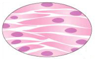
Smooth muscles are made up of numerous extended muscle fibers which considerably vary in size. The smooth muscle fibers are in general shorter than skeletal muscle fibers. Each smooth muscle fiber is a single cell surrounded by sarcolemma. The cells are fusiform in shape that means spindle shaped having a wider middle portion called belly and the tapering ends. Each cell contains a single, oval nucleus placed at the center of the belly. The muscle fibers are arranged in such a way that the wider middle portion of one fibre is placed in-between the narrowing ends of adjacent fibers. Each fiber is externally covered by a basement membrane like structure which separates the fibers from each other, some smooth muscles, the basement membrane is discontinuous at places where sarcolemma of adjacent cells fuse together to form 'tight junctions' or low electrical resistance bridges comparable to the inserted discs of cardiac muscle.

Smooth muscle of intestine
Types of smooth muscles :
Smooth muscles into two types—single unit smooth muscle and multi-unit smooth muscle which differ in various features. Now let us go for some discussion:
Single unit smooth muscle :
It is commonly called visceral smooth muscle because it is found in the walls of hollow visceral organs like gut, gall bladder, urinary bladder, ureter, uterus etc. In this type of muscle, the muscle fibers are usually arranged in sheets or bundles and they remain unified through low electrical resistance bridges forming a functional syncytium. Thus, when one fiber is excited, the excitation or impulse spreads to the neighboring fibers and as a result large number of muscle fibers contact simultaneously as if they belong to a single unit. For this reason, this type of muscle is known as single unit smooth muscle. Due to the presence of functional connection between the fibers, each fiber is not innervated by a distinct nerve ending. The muscle fibers show pacemaker like activity and they generate action potentials when stretched. So they contract spontaneously in response to stretch even when de-enervated. However, the nerves may modify their shrinkages.
Multi-unit smooth muscle :
This type of smooth muscle found in ciliary and iris muscles of eye, piloerector muscles of skin and in larger blood vessels. This type of muscle composed of distinct muscle fibers without low electrical resistance bridges in between them. Each fiber is completely covered by the basement membrane and remains separated from the neighboring fibers. Each fiber is innervated by a separate nerve ending and operates independently of the others. Since, the individual fibers behave as separate units, this type of smooth muscle is called multi-unit smooth muscle. These muscles do not show pacemaker activity and they do not contract instinctively in response to stretch. Muscle fibers are activated only when stimulated by nervous or chemical agents like hormones.
Contraction and relaxation of smooth muscles

Contraction and relaxation of smooth muscle